Aortic atlas
Below is my official aortic atlas, in which I have compiled information about the anatomy of the aorta. I hope you find it useful, and, as always: let me know if you have any questions.
Click the chapter titles to the left (at the top on smaller screens) to learn about different aspects of the aorta.
Anatomy of the thoracic aorta
Anatomy of the abdominal aorta
Abdominal aortic aneurysms
Endovascular aneurysm repair
Aortic coarctation
Anatomy of the thoracic aorta
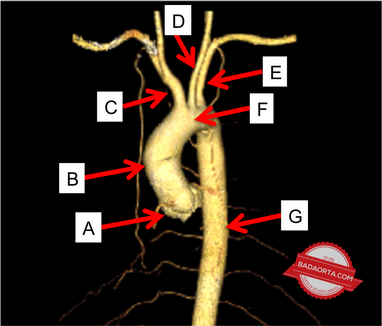
A. Aortic root
- This is the section of the aorta starting at the aortic valve and extending the the sinotubular junction which is the transition between the aortic root and the ascending aorta
- The aortic root contains the origins of both the left and right main coronary arteries which provide blood supply to the heart muscle (myocardium)
- The aortic root billows out into 3 pouches, called sinus of valsava
B. Ascending aorta
- The ascending thoracic aorta starts at the sinotubular junction and ends just before the start of the innominate artery
- Many aortic dissections begin in the ascending aorta (Stanford Type A)
C. Innominate artery (also called the brachiocephalic artery)
- This is the first branch off the aorta
- The innominate artery branches into the right subclavian artery and right common carotid artery
- A “bovine arch” refers to a common variant of anatomy where the left common artery originates off the innominate artery instead of directly off the aortic arch
D. Left common carotid artery
- The left common carotid artery is the second branch off the aorta and is located in the aortic arch
- A “bovine arch” refers to a common anatomic variant where the left common carotid artery originates off the innominate artery instead of directly off the aortic arch
- The left common carotid artery supplies blood to the left side of the head and brain
E. Left subclavian artery
- The left subclavian artery is the third branch off the aorta and is located in the aortic arch
- The left subclavian artery supplies blood to the left arm
F. Aortic arch
- The aortic arch is the third segment of the aorta after the aortic root and ascending thoracic aorta
- It gives rise to the innominate artery, the left common carotid artery and the left subclavian artery
G. Descending thoracic aorta
- The descending thoracic aorta begins immediately after the left subclavian artery and continues until the origin of the celiac artery in the abdomen
- Many aortic dissections originate in the proximal descending thoracic, just beyond the left subclavian artery (Stanford Type B aortic dissection)
Anatomy of the abdominal aorta
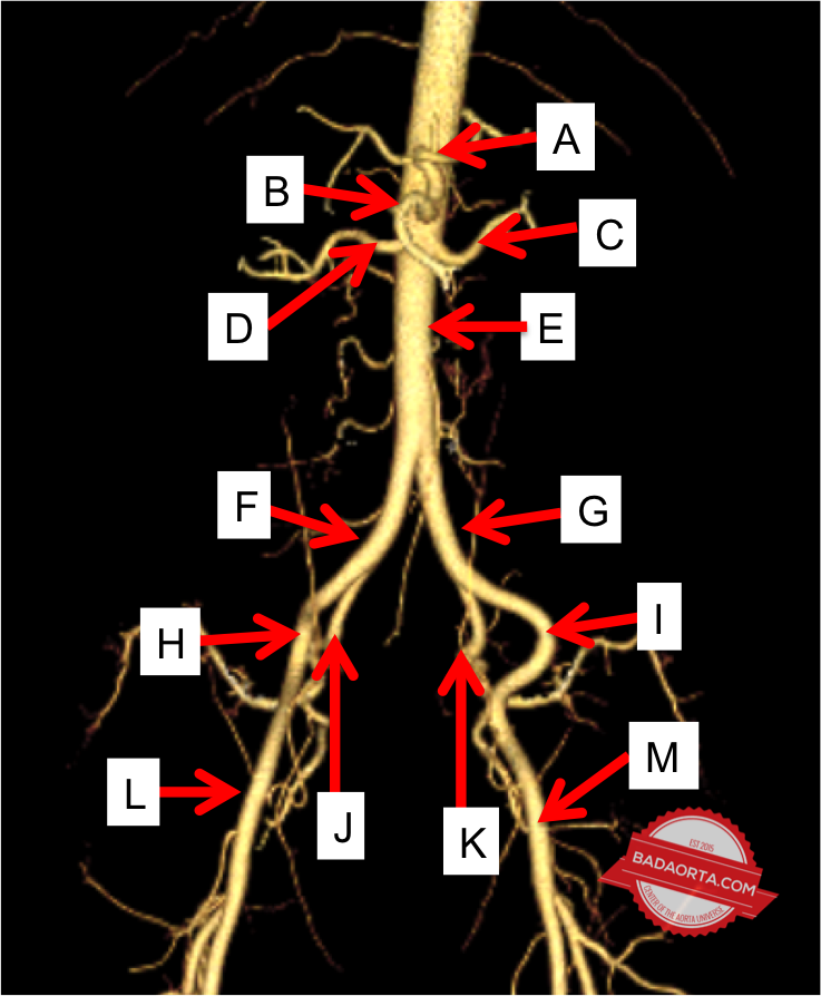
A. Celiac artery
- This is the first branch off the aorta below the diaphragm
- Also called the celiac trunk
- Branches into the splenic artery and the hepatic artery
B. Superior mesenteric artery
- The second branch off the aorta below the diaphragm
- Provides blood flow to the intestines and colon
C. Left renal artery
- Provides blood flow to the left kidney
D. Right renal artery
- Provides blood flow to the right kidney
E. Infrarenal abdominal aorta
- Predisposed to developing an abdominal aortic aneurysm
- Gives rise to the inferior mesenteric artery
F. Right common iliac artery
- Provides blood flow to the right leg
- Can develop an aneurysm or blockage
G. Left common iliac artery
- Provides blood flow to the left leg
- Can develop an aneurysm or blockage
H. Right external iliac artery
I. Left external iliac artery
J. Right internal iliac artery
- Also called the hypogastric artery
K. Left internal iliac artery
- Also called the hypogastric artery
M. Right common femoral artery
N. Left common femoral artery
Abdominal aortic aneurysms
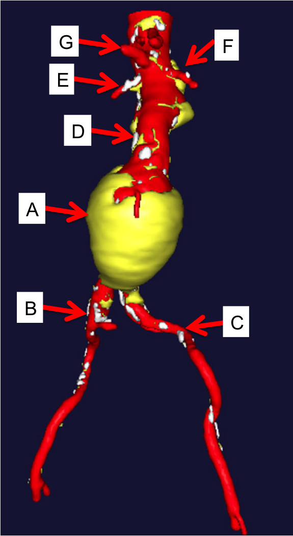
A. Abdominal aortic aneurysm (AAA)
- The segment of the aorta below the renal arteries and above the aortic bifurcation is a common location for aortic aneurysms to develop
- The maximal width – not length – of the abdominal aortic segment is what determines risk of rupture
- A maximal diameter of 5cm at the aortic segment is a trigger for concern and a point at which to begin the discussion for treatment
B. Right common iliac artery
- The aortic bifurcation represents the anatomic end of the aorta
- The aorta divides into the right and left iliac arteries at the aortic bifurcation
- The right common iliac artery represents the beginning of blood supply to the right leg
- The common iliac artery (right or left) can become aneurysmal
C. Left common iliac artery
- The aortic bifurcation represents the anatomic end of the aorta
- The aorta divides into the left and right iliac arteries at the aortic bifurcation
- The left common iliac artery represents the beginning of the blood supply to the left leg
- The common iliac artery (right or left) can become aneurysmal
D. Abdominal aorta
- The abdominal aorta is the segment of the aorta below the diaphragm where the aorta enters the abdomen
- The normal diameter of the aorta varies by patient and is related to height, weight and genetics
- The abdominal aorta contains several branches to the abdominal organs
- The abdominal aorta is subject to several different types of diseases including aneurysms (expansion) or occlusion (blockage)
E. Right renal artery
- The right renal artery is the blood supply to the right kidney
- Some people are predisposed to an accessory renal artery (extra renal artery) which does not have any significant consequences
F. Left renal artery
- The right renal artery is the blood supply to the right kidney
- Some people are predisposed to an accessory renal artery (extra renal artery) which does not have any significant consequences
G. Superior mesenteric artery
- The superior mesenteric artery provides blood supply to both the small and large intestines
Endovascular aneurysm repair
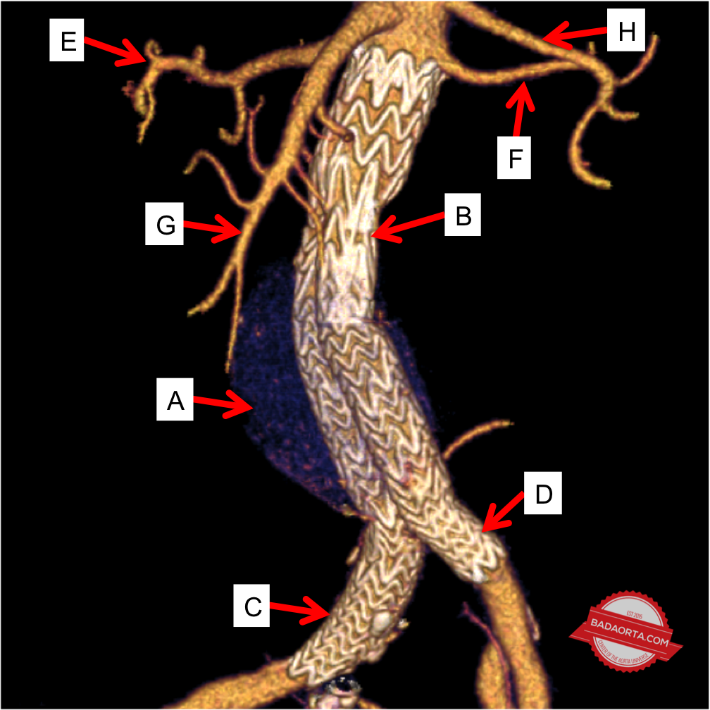
A. Abdominal aortic aneurysm (AAA)
- An abdominal aortic aneurysm (AAA) usually develops in the segment of the abdominal aorta below the renal arteries and above the aortic bifurcation
- When an abdominal aortic aneurysm is treated with an endovascular aortic stent-graft, the aneurysm sac stays intact, but all of the blood flow in the aneurysm is constrained within the aortic stent
B. Endovascular Abdominal aortic stent-graft (EVAR)
- Also called “EVAR”, which stands for endovascular aneurysm repair
- An endovascular aortic stent-graft is a medical device which is inserted inside the aortic aneurysm using special endovascular techniques and x-ray guidance
- The abdominal aortic stent-graft stays in the aorta permanently and prevents the aortic aneurysm from growing/expanding or rupturing
C. Right limb of abdominal aortic stent-graft
- EVAR, which stands for endovascular aneurysm repair, includes extension of the abdominal aortic stent into both iliac arteries
- These bifurcated devices are necessary in order to preserve blood supply to the lower extremities and prevent flowing blood from entering the aneurysm sac
D. Left limb of the abdominal aortic stent-graft
- EVAR, which stands for endovascular aneurysm repair, includes extension of the abdominal aortic stent into both iliac arteries
- These bifurcated devices are necessary in order to preserve blood supply to the lower extremities and prevent flowing blood from entering the aneurysm sac
E. Right renal artery
- The right renal artery supplies blood to the right kidney
- Most abdominal aortic stent-grafts do not obstruct or block blood flow to the kidney arteries
- There are some custom aortic stent-grafts which have branches and allow blood flow to the renal arteries
F. Left renal artery
- The right renal artery supplies blood to the right kidney
- Most abdominal aortic stent-grafts do not obstruct or block blood flow to the kidney arteries
- There are some custom aortic stent-grafts which have branches and allow blood flow to the renal arteries
G. Superior mesenteric artery
- The superior mesenteric artery supplies blood to the small and large intestines
- In certain circumstances, the abdominal aortic aneurysm extends above the renal arteries to the level of the superior mesenteric artery
H. Celiac artery (Celiac trunk)
- The celiac artery (celiac trunk) is the first branch off the aorta below the diaphragm
- The celiac artery supplies blood to the stomach, liver and spleen
Aortic coarctation
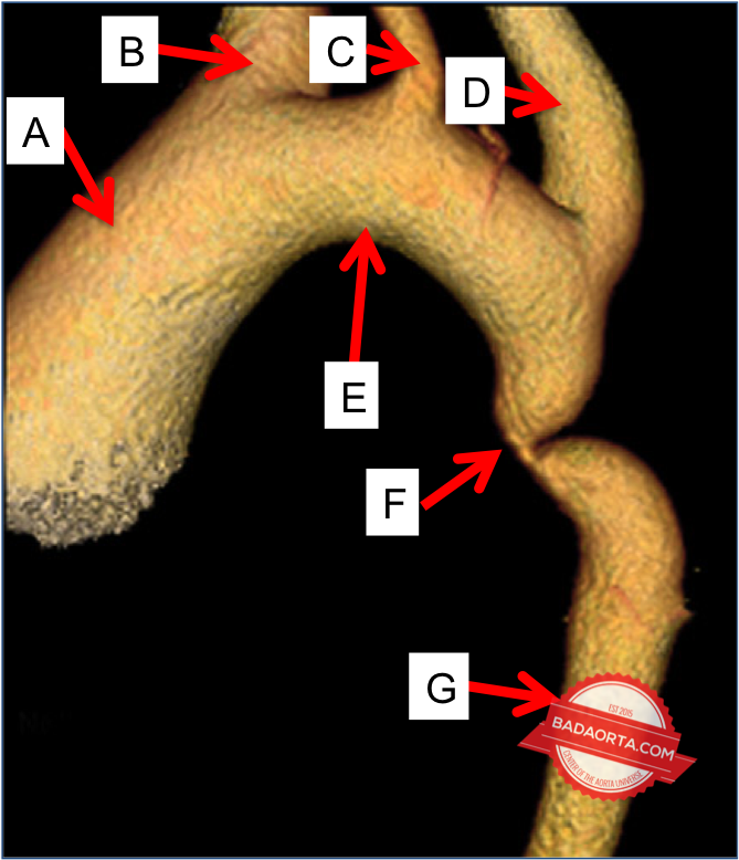
A. Ascending aorta
- The ascending aorta is just above the aortic root where the aorta interfaces with the aortic valve and left ventricle of the heart
- The ascending aorta can be dilated or enlarged in aortic coarctation due to an association with bicuspid aortic valve syndrome in patients with aortic coaractation
B. Innominate artery
- The first branch off the aorta in the aortic arch
- Supplies blood to the right arm and right side of the face and brain
C. Left common carotid artery
- The second branch off the aorta in the aortic arch
- Supplies blood to the left side of the face and brain
D. Left subclavian artery
- The third branch off the aorta in the aortic arch
- Supplies blood to the left arm
- Occasionally, the left subclavian artery is intimately involved in aortic coarctation process
E. Aortic arch
- The aortic arch is the third anatomic segment following the aortic root and ascending aorta
- Gives rise to the great vessels which supply blood to the upper extremities, head and brain
F. Aortic coarctation
- A congenital (from birth) narrowing of the aorta which develops just beyond the origin of the left subclavian artery in the proximal descending thoracic aorta
- Sometimes aortic coarctation is diagnosed shortly after birth and repaired, yet can occasionally be diagnosed and treated later in life as a teenager or adult
G. Descending thoracic aorta
- The descending thoracic aorta is the fourth anatomic segment of the aorta following the aortic root, ascending aorta and aortic arch