Medical Arc de Triomphe (or Why your Aortic Arch is Unique)
Introduction
The Arc de Triomphe de l’Étoile in Paris was completed in 1808, and stands as both an architectural monument as well as a source of French national pride.

Did you know that the human Aorta has its own “Arc de Triomphe”?
Yes, the Aorta is divided into several distinct anatomic segments including: aortic root, ascending aorta, aortic arch, descending thoracic aorta and abdominal aorta.
Each of these segments are predisposed to different types of stresses and strains and associated problems which we will discuss.
Anatomy of the Aortic Arch
The aortic arch is a medical marvel. Let me explain. As blood is ejected from the left ventricle of the heart, it passes quickly through the aortic root, past the left and right main coronary arteries and into the ascending aorta. Next, blood enters the aortic arch and goes in several directions. Most of the blood passes around the aortic arch and into the descending thoracic aorta. Simultaneously, blood enters all three vessels (innominate artery, left common carotid artery and left subclavian artery) which provides blood to the upper extremities, face and brain.
Think about that for a moment: all the blood that our brain requires passes through the aortic arch. Just like the Arc de Triomphe, this segment of the aorta is the crown and jewel due to the central anatomic and physiologic role it plays with supplying blood to the brain.
Let’s look a little closer at the aortic arch and its anatomy.
Great vessels of the aortic arch
The “great vessels” entail the branches originating at the apex of the aortic arch. As the ascending aorta sweeps around, the aorta transitions into the aortic arch. This allows the aorta to curve around and transition to a segment (descending thoracic aorta) which runs the length of the torso and provides blood to our internal organs. If you recall, we discussed the embryology of the aortic arch in a previous post.
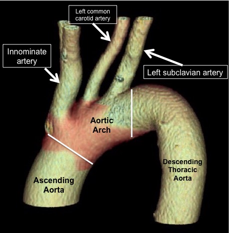
As seen in this CT scan reconstruction, the aortic arch begins with the origin of the first branch, called the innominate artery. This artery provides blood flow to both the right arm through the right subclavian artery and also to the right side of the face and brain. The next branch in the arch is the left common carotid artery which supplies the left side of the face and brain. The third branch is the left subclavian artery supplying blood to the left arm.
Anatomic variations of the aortic arch
Most importantly, there are anatomic variations to the great vessel anatomy. A bovine arch describes a situation where the left common carotid artery originates not from the Aorta, but from the innominate artery. These patients have only 2 great vessels. This configuration doesn’t have any affects on blood flow and most people with this configuration don’t even know they have this variant. It occurs in about 10% of the population.
Another anatomic variation is having 4 vessels (instead of the normal 3) coming off the aortic arch. One scenario is having the left vertebral artery (usually a branch off the left subclavian artery) originate directly off the aortic arch. This artery supplies the posterior aspect of the brain. Another scenario is having an aberrant right subclavian artery. This is a congenital disorder where the artery to the right arm (right subclavian artery) originates off the distal aortic arch instead of the innominate artery. This means that the artery to the right arm passes behind the aortic arch.
Dysphagia lusoria
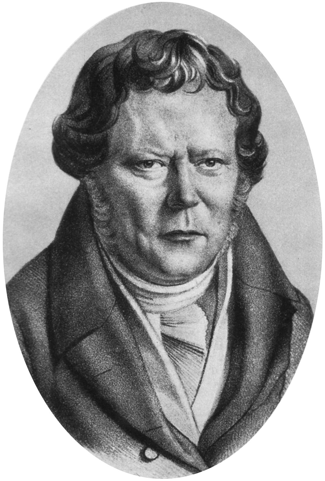
A little know fact is that Johann Heinrich Ferdinand von Autenrieth, a german physician living in the early 1800’s, described the medical phenomenon called dysphagia lusoria. This is the medical condition where the esophagus is compressed by the aberrant right subclavian artery passing behind the the aortic arch as a result of this artery being the 4th (and last) branch of the aortic arch. Dysphagia lusoria is associated with trouble swallowing as a result of the compression of the esophagus. This is a rare condition, but when it occurs, the condition can be resolved with surgery.
In conclusion
In summary, the aortic arch is a medical marvel due to it’s integral nature of providing blood to the upper extremities, face and brain. You can imagine the sometimes disastrous consequences of alterations and disruption of the great vessels. The aortic arch is a complex structure. In future posts, we will discuss the diseases and problems that can occur in the aortic arch and also surgical and endovascular treatments. Hybrid procedures, aortic stents and open aortic surgery are some of the options.
Question
What are your thoughts?
Do you agree that the Aortic Arch is a Medical Arch de Triomphe?
Was this post informative?
Subscribe to my newsletter to learn more about the aorta, its diseases, and how to treat them.
Comments
Share your thoughts below — I try to get back to as many comments as possible.